
As farm vets out on the road, we carry our office and pharmacy with us in our vehicles. One bit of kit I will never be without is my ultrasound scanner. I use it nearly every day and while we tend to use it primarily for pregnancy diagnosis in cattle, there are also several other times it can be used as a diagnostic tool.
Ultrasound scanners were not always affordable or widely used by farm vets in practice, vets used to rely more heavily on manual palpation techniques to assess the fertility status of the uterus and ovaries. While this is a technique that we still use there is nothing better than being able to visualise what you can feel, especially when you see a little foetus wriggling around inside. Often more accurate diagnosis can be made too, especially as you can see details such as the foetal heartbeat or the presence of twins.
Another use is to sex the foetus. Once the foetus reaches 70 days it is possible to do so. We look for the genital tubercle which is a structure found between the hindlegs in all younger pregnancies, then as the foetus develops it moves either towards the tail or the umbilical cord to develop into the female or male genitals respectively. It is a bilobed oval structure that appears hyperechoic – bright white – on the scan, often appearing as two little white lines.
It is also useful to be able to visualise something that feels abnormal. For example, last week I diagnosed an ovarian tumour in a cow, who had not been able to get in calf. The ovary felt huge and using the scanner you could see how abnormal the tissue consistency was.
The ultrasound scanner can also be used externally on animals to visualise different areas. Our probes are designed to use rectally, so it is important when using them on skin to prep the area carefully to ensure the best picture is achieved, and to do so a close contact is required. We prep the area by clipping the hair and we need to achieve a close shave. Then I soak the skin by applying warm water and then apply liberal amounts of ultrasound gel, giving it five minutes to soak in before scanning. A gentle amount of pressure is then applied to maintain a good contact. The depth and frequency of the ultrasound waves can be adjusted depending on what you are scanning and how far away it may be or how much detail you wish to see.
Ultrasound can be used to assess udder health, especially if there is an unexplained enlargement of the gland which could be due to a haematoma, abscess or oedema. It can also be used to examine the teat, to assess the inner structure and confirm diagnosis of fibrosis or stenosis of the teat canal.
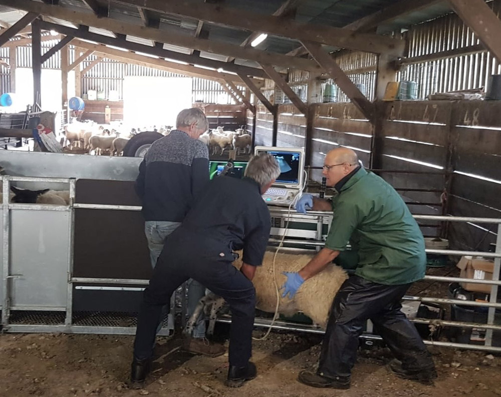
The scanner can be used to assess both the thoracic cavity, and the abdominal cavity in sheep and cattle. It is useful to assess the heart and lungs where clinical examination raises suspicion of certain conditions including pneumonia, lung lobe consolidation, pleural effusion, septic pericarditis or OPA – ovine pulmonary adenocarcinoma – in sheep. Abdominal scanning can help assess structures such as the liver, kidney, reticulum and spleen.
When calves present with navel swellings the scanner can help diagnose an abscess as well as confirming the presence or absence of a hernia, a result which would dictate the treatment path of choice. I have even found it a useful tool for assessing the fertility status of bulls, where lesions are found in the testicles. So, while we can’t fit our patients into a small animal diagnostic centre with all the fancy equipment at hand, it doesn’t mean that we can’t still tell a lot using what we have in the back of the car!
by Alice Miller BVSC DBR MRCVS -Friars Moor Livestock Health


Leave a Reply