
by Lynn Broom.
Longmead Veterinary Practice.
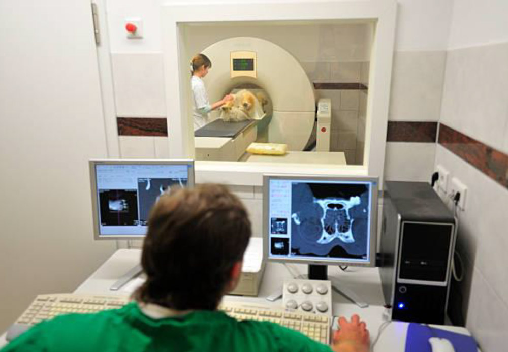
CT scans – previously known as CAT scans – are images created by computed tomography and are a series of x-rays, interpreted by a computer, to give a detailed view of the inside of the body as a cross-section. A 3D image can be produced so that the area of interest can be further assessed.
A CT scanner is a complex and expensive piece of equipment which needs its own dust-free environment, controlled temperature and a steady level of humidity, and is housed within a room entirely lined with lead to minimise spread of x-rays.
Traditional x-rays primarily show bony changes, although differences in soft tissue densities will also be visible. However, they do not give the detail required for smaller areas of soft tissue or where different soft tissue structures have the same density. Soft tissue under or within bone, such as the brain, cannot be assessed on x-rays because bone obscures this.
Ultrasound scanning allows assessment of soft tissue structures and is useful to assess abdominal structures and soft tissue structures. Again it is limited by the amount of detail it can provide and ultrasound cannot ‘see’ through bones or air, which limits its use in the chest, the skull and the spine.
The CT scanner produces multiple closely placed images which the computer interprets and small fine structures can be identified which would otherwise be unseen on an x-ray or ultrasound image. Contrast medium injected in to the veinous system allows specific imaging of areas otherwise unseen or poorly defined.
CT scans provide excellent images of bones to allow, for instance, assessment of complex fractures prior to surgery, and the lungs to help identify areas of damage due to, for example, inhaled foreign bodies or tumours. Using a contrast medium the urinary system is also clearly seen and can identify obstructions within, for example, the ureters, which carry urine from the kidneys to the bladder, which can be difficult to properly assess with other imaging media.
Foreign bodies can also be visualised in other inaccessible areas which are difficult to x-ray or ultrasound scan such as behind the eye. Tumours within nasal chambers can be difficult to assess and CT scans are very useful in imaging this area.
CT scans are also good at assessing soft tissues within bony structures such as the skull and spine to allow assessment of the brain to identify brain tumours and bleeds. The spinal cord can be assessed for lesions and assessment of slipped discs, using contrast media where appropriate.
Other structures within the chest, abdomen and intestines are also identifiable in more precise detail than x-rays and ultrasound allowing, for instance, precise diagnosis before surgery is performed.
MRI – magnetic resonance imaging – scans are more precise for certain structures such as nerves, fine brain detail and muscles, although adequate views of these structures can be seen on CT scan. A clinical decision has to be made as to which type of scan is more appropriate to the condition being investigated. Neuromas, which are tumours of nerves, are visible on CT scans. MRI scanners are considerably more expensive, require even more specific environmental controls and the scans themselves take much longer.
CT scans are an excellent addition to our repertoire of diagnostic options, especially where further detail is required which cannot be provided by x-rays or ultrasound scans and where cost prohibits MRI scanning.
CT scans extra tool in vet’s armoury

Related Articles

Have your say over new rules for dogs in Dorset’s public spaces
DOG owners and others are being urged to have their say on rules for…

Five-year ban after woman filmed repeatedly beating dog
A WOMAN who was caught on camera repeatedly mistreating two defenceless dogs has been…

Secure dog walking area planned for Dorset village
DOG walkers in a village near Yetminster could eventually…


Leave a Reply